x

Key Research Focus

  • Image Segmentation & Feature Extraction 
  • Multi-modality image registration
  • 3D Reconstruction and anatomical model generation
  • AI & DL for Image analysis 
  • Predictive Imaging

Projects

Intra-operative Fluoroscopic Image Registration

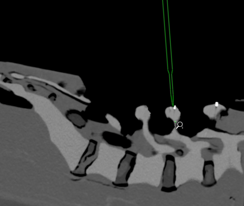
Computer-aided surgery (CAS), especially in spinal procedures, benefits greatly from advancements in imaging, navigation, and robotics. This research develops novel intraoperative registration techniques for fluoroscopic images to enhance spatial awareness and real-time guidance. By integrating tracking data with registered images, the system ensures accurate instrument localization, precise implant placement, and improved navigation—crucial for minimizing complications and enhancing outcomes in minimally invasive surgeries.

Research Focus:

  • Spatio2-frequency Image Enhancement
  • Image Distortion Correction
  • Feature Extraction
  • Pose Identification
  • Image Registration

Pre-operative 3D volumetric data to Intra-op 2D fluoroscopic image registration for Open and Minimally Invasive Surgeries

The process of 2D-3D registration entails correlating anatomical features or landmarks between the 2D and the 3D images. Key steps involve obtaining high-resolution preoperative scans, identifying landmarks in both image sets, and determining a transformation to align them. This alignment enhances surgical guidance by superimposing preoperative anatomy onto the live fluoroscopic view, facilitating precise navigation. The integration of data leads to comprehensive visualization, improved spatial orientation and enhanced anatomical details thereby improving  surgical accuracy and safety, benefiting both open and minimally invasive procedures.


Research Focus:

  • Feature mapping
  • Multi-modality registration
  • Digital reconstruction of radiographs
  • 3D visualization

GUI-Based Pedicle Screw Planning

Image-based intraoperative planning improves surgical accuracy and reduces complications, especially in procedures like pedicle screw insertion. Since intraoperative CT is often impractical, planning on fluoroscopic images is a more efficient alternative. This research aims to develop a GUI-based platform that allows surgeons to plan screw placement using simulated 3D screw projections on fluoroscopic images. The system uses 2D vertebral segmentation for screw initialization and positioning, offering a practical, time-saving tool to support surgeons during spinal surgeries.


Research Focus:

  • Projective Correspondence
  • Vertebral segmentation in 2D utilizing DL
  • Synchronous Planning
  • GUI Feature set

Deep Learning-Based Spinal Anatomy Detection in X-Rays

Classical direct rendering from CT data requires numerous images, increasing radiation exposure and limiting accessibility in low-resource settings. This project explores inverse rendering to reconstruct 3D volumes from just two 2D X-ray images, reducing both cost and radiation. We propose a deep learning pipeline using
neural style transfer—adapted from artistic image processing—to medical X-rays and digitally reconstructed radiographs (DRRs). The goal is to enable accurate 3D reconstruction and 2D-to-3D registration, advancing affordable and low-dose imaging solutions

Research Focus:

  • Neural Style Transfer
  • DL based inverse rendering
  • 3D Reconstruction
  • Virtual Correspondence

Fluoro-CT Spline Registration Utilizing Pose Based Convolutional Neural Network

In order to improve surgical navigation accuracy, this work proposes a mechanism for matching 3D CT scans with 2D X-ray pictures. To generate a 3D volume, CT slices were first obtained and processed. Annotation and model training then took place. Predicted regions of interest were used to clip the CT volume, and digitally recreated radiographs (DRRs) were produced for surgical planning. For image registration, rigid transformation and gradient-normalized cross-correlation were used, and different optimization strategies were investigated for effectiveness. Both qualitative and quantitative studies were used to evaluate the methodology's efficacy, with a focus on registration accuracy and computing efficiency.

Predicting Occluded Fiducials with GANs Using Pose Priors

In interventional radiology and surgery, C-arm systems with calibration drums aid in efficient imaging. However, occlusion often hides some calibration fiducials in X-ray images. To address this, we use pix2pix GANs to predict and generate synthetic images with complete fiducial sets. Pix2pix GAN, a deep learning model requiring minimal paired training data, consists of a U-Net generator and PatchGAN discriminator. The U-Net captures and reconstructs image features, while PatchGAN evaluates image realism at the patch level, enabling accurate and efficient fiducial prediction for improved 2D-2D registration.

Optimizing Pedicle Screw Placement Parameters for IGSS

Image-guided spine surgery (IGSS) requires high precision in pedicle screw placement. This study presents a novel approach using adaptive parameter tuning to improve placement accuracy. Key parameters—such as sigma (Laplacian of Gaussian), contrast adjustment (x), and clustering metrics (epsilon, minPts)—were optimized through iterative testing and reprojection error analysis. Linear regression with the CVX library was used to identify configurations that minimized error. The model dynamically adapts parameters based on real-time image contrast, enhancing accuracy and efficiency in IGSS procedures.

3D Ultrasound reconstruction from 2D scans

Ultrasound reconstruction from 2D refers to the process of generating three-dimensional images from a series of two-dimensional ultrasound scans.

Publications

Aparna Purayath,  Vivek Maik, Abhilash C, Manojkumar Lakshmanan, and Mohanasankar Sivaprakasam.

Vivek Maik, Aparna Purayath, Durga R, Manojkumar Lakshmanan, and Mohanasankar Sivaprakasam. 

Yaswantha Rao P, Gaurisankar S, Durga R, Aparna Purayath, Vivek Maik, Manojkumar Lakshmanan, and Mohanasankar Sivaprakasam. 

Our Team